Treating the Dental Patient with a Developmental Disorder
 Scully’s Medical Problems in Dentistry, 7th Edition
 Rialzo del seno mascellare (T. class) Italian with English Subtitles
 Mosby‘s Review Questions for the National Board Dental Hygiene Examination
 Immediate Loading of Dental Implants: Theory and Clinical Practice
 Dislocation of the Temporomandibular Joint: A Guide to Diagnosis and Management
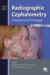 Radiographic Cephalometry: From Basics to 3-D Imaging, 2nd Edition

Description
Radiographic cephalometry has been one of the most important diagnostic tools in orthodontics, since its introduction in the early 1930s by Broadbent in the United States and Hofrath in Germany. Generations of orthodontists have relied on the interpretation of these images for their diagnosis and treatment planning as well as for the long-term follow-up of growth and treatment results. Also in the planning for surgical orthodontic corrections of jaw discrepancies, lateral and antero-posterior cephalograms have been valuable tools. For these purposes numerous cephalometric analyses are available. However, a major drawback of the existing technique is that it renders only a two-dimensional representation of a three-dimensional structure.
With “Three-Dimensional Cephalometry – A Color Atlas and Manual” by the authors Swennen, Schutyser and Hausamen you have an exciting book in your hands. It shows you how the head can be analysed in three dimensions with the aid of 3D-cephalometry. Of course, at the moment the technique is not available in every orthodontic office around the corner.
| Author(s): Gwen R.J. Swennen - Filip Schutyser |
|
English | 2006 | ISBN: 9783540254409 | 367 pages | PDF | 25 MB |