Clinical Practice of the Dental Hygienist, 12th Edition & Active Learning Workbook
 Essential Dental Therapeutics
 Lippincott Williams & Wilkins’ Comprehensive Dental Assisting (with workbook)
 Panoramic Radiology: Seminars on Maxillofacial Imaging and Interpretation
 MCQs for Oral Radiology: Principles and Interpretation
 Contemporary Cephalometric Radiography
 The Dentists Quick Guide to Medical Conditions
 Cone Beam Volumetric Imaging in Dental, Oral and Maxillofacial Medicine
 Failure in the Restored Dentition: Management and Treatment
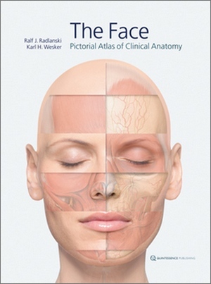
Description
All surgical disciplines that deal with the human face must rely on an exact knowledge of its highly complex topographic-anatomical relationships. This atlas provides a unique contribution to the study of facial anatomy in which readers are allowed to explore deeper into the anatomy as the layers are stripped away, one by one. This book features elaborate composite illustrations created in layers, starting from the skeleton and working out to the surface of the skin. CT scans are used for the skeletal template, MRI series depict the inner layers of the facial region, angiographs report the anatomy of the blood vessels, and photographs of anatomical specimens as well as a living model fill out the rest. The final products are anatomical images of hyper 3D clarity and arresting beauty. Significant landmarks, anatomical details, and clinically relevant groupings of nerves and blood vessels are the primary focus of each image series to preserve clinical relevance. Accompanying text and figure legends emphasize key points, but the focus is always on the vivid anatomical images.
Scanned Searchable Copy
| Authors: Ralf J. Radlanski , Karl H. Wesker |
|
English | 2012 | ISBN: 9781850972143 | 359 pages | PDF | 272 MB |