 Prosthetic Treatment of the Edentulous Patient, 5th Edition
 Cephalometry in Orthodontics 2D and 3D
 Orofacial Pain: A Clinicians Guide
 Scully’s Medical Problems in Dentistry, 7th Edition
 Dentistry and the Pregnant Patient
 Review Questions for Dentistry
 High-Strength Ceramics: Interdisciplinary Perspectives
 The Root Canal Biofilm
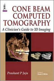 Cone Beam Computed Tomography: A Clinician's Guide to 3d Imaging
Description
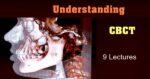
This course will move your 3D diagnostic skills in the area of implant dentistry to the next level. Review the systems and anatomy viewed on a CBCT scan one structure at a time using radiographic images and graphic representations. Understand clinical relevance of each anatomic structure along with how variations of these structures can affect the your diagnosis and treatment plan.
Part-1
- Understanding CBCT Technology
- Radiographic anatomy of the maxilla, mandible and osteomeatal complex
- Alveolar process morphology variation and their effect on implant analysis
Part-2
- Anatomy related errors
- The process of disuse atrophy and how it changes the position of the crest of the ridge
- Evaluating the implant site with the occlusion and function in mind
- The process of osteointegration and how this relates to radiographic evaluation
- Treatment planning around pathology
Part-3
- Practical CBCT
Presenter: Dania Tamimi
3 Videos - Duration 4 hours and 20 minutes - Files size 700 mb